The BrainStem Bundle Tool (BSBT) instantly segments eight critical brainstem pathways from a standard diffusion MRI scan, giving clinicians a quantitative map of the region that controls breathing, heart rate, and consciousness. By feeding raw images into a deep‑learning network, BSBT eliminates manual tracing, speeds up analysis, and opens new diagnostic possibilities for you and your patients.
How BSBT Transforms Brainstem Imaging
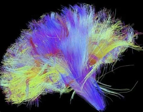
Traditional brainstem imaging has been hampered by tiny, densely packed fibers that blur together on conventional scans. BSBT tackles that problem head‑on. It ingests diffusion‑weighted images, runs them through a trained neural net, and outputs a clean segmentation of each bundle in seconds. The result is a clear, quantitative view that was previously impossible to obtain.
Deep‑Learning Engine Behind the Segmentation
The algorithm was trained on expertly labeled datasets, teaching it to recognize the unique diffusion signatures of each pathway. When you upload a scan, the model filters out cerebrospinal fluid noise and motion artifacts, then labels the eight major bundles:
- Corticospinal tract
- Medial lemniscus
- Dorsal longitudinal fasciculus
- Other five key brainstem pathways
Because the network learns directly from the data, it adapts to variations in scanner hardware without needing specialized equipment.
Clinical Impact Across Neurological Disorders
BSBT’s quantitative maps give doctors a new way to track disease‑related changes. In early tests, the tool highlighted distinct structural alterations in patients with Parkinson’s disease, multiple sclerosis, and traumatic brain injury. It also detected subtle shifts in Alzheimer’s cohorts, suggesting a broad biomarker potential.
Case Study: Tracking Recovery in a Coma Patient
One retrospective analysis followed a patient who emerged from a seven‑month coma. BSBT traced a gradual restoration of specific brainstem bundles, mirroring the patient’s clinical improvement. That kind of longitudinal insight lets you see recovery patterns that were previously hidden.
Practitioner Perspective
Neuroradiologists say the tool changes how they describe lesions. Instead of vague phrases like “a subtle hyperintensity in the pons,” they can now provide precise, bundle‑by‑bundle measurements. The software runs on a standard workstation, so you don’t need a high‑end GPU cluster to benefit.
Future Directions and Adoption
The developers envision BSBT becoming a routine part of radiology pipelines. Imagine receiving an automated bundle report alongside the standard scan—no extra steps, just richer data. As more hospitals adopt the tool, community benchmarks will sharpen its accuracy even further.
Integration Into Clinical Workflows
Because BSBT works with any diffusion MRI sequence, it sidesteps the need for proprietary protocols. Radiology departments can plug it into existing PACS systems, and clinicians can start using quantitative brainstem metrics today. If you’re looking to enhance diagnostic confidence, BSBT offers a ready‑to‑use solution.